ScoutXcan

MULTI-ANGLE DIGITAL 2D X-RAY SYSTEM

Visualise a core’s internal structure with X-ray Imaging

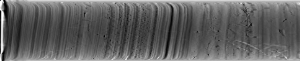
Geotek’s ScoutXcan is an affordable X-ray scanning system which has the capability to produce 2D multi-angle X-ray radiographs from linear and rotational 2D imaging. Using Geotek’s geoscience-ready laminography acquisition method, the ScoutXcan can successfully identify pseudo 3D information from rock and sediment core samples without the need for full 3D CT. This makes it the ideal system is ideal for customers on a budget and that do not require full 3D CT imaging.

CABINET SAFE – No need for a lead-lined room!

Mobile Laboratory, Onshore or Offshore Worldwide
Istituto Nazionale di Oceanografia e di Geofisica Sperimentale (OGS), Trieste
Arctic University of Norway (UiT), Tromso

How it works

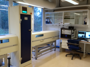
Samples are loaded horizontally into one of the cabinet arms inside of an X-ray tube and centralised by wirelessly controlled motorised arms. The core sample translates through the centre cabinet past a fixed source and detector position that the user has selected, and can be rotated under computer control to examine internal structures at various angles to give the user pseudo 3D information.

DATA ACQUISITION & SOFTWARE

The user is able to scan their core samples using various acquisition modes which may be selected prior to scanning: 2D radiography and laminography

  • Geotek X-ray acquisition software – allows the operator to set-up their scan parameters and acquire data with real-time visualisation.
  • Geotek GQuickView software – allows the user to view and manipulate the X-ray radiographs and CT slabs.

previous arrow
6.0 Software
Geotek X-ray acquisition software
Geotek XCT Software
GQuickView
Geotek GQuickView Software
next arrow

ACQUISITION MODES

The user is able to scan their core samples using various acquisition modes which may be selected prior to scanning; 2D radiography and laminography

  • 2D Radiography
    • Multi-angle 2D transmission images with software corrections to produce virtual slab

  • 3D Laminography
    • Quasi-3D imaging technique using tomographic reconstruction to reconstruct planes within the core

  • Specifications

    • X-ray source: 65 W Thermo Kevex 130 kV Microfocus
    • Voltage Range: 45 kV to 130 kV
    • X-ray Spot Size Range: 7 μm (at 4W) – 100 μm (at 65 W)
    • Detector: 1920 x 1536 pixel flat panel detector
    • Voxel/Pixel Resolution Range: 30 µm to 350 µm
    • Size (L x W x H mm): 130 kV – 4997 x 700 x 2022
    • Weight: 130 kV – c. 1200 kg
    • Utilities: 100 V to 240 V, single phase, 50-60 Hz
    • Cooling: None
    • Core/Plug Dimensions: Diameter – 2.5 cm to 15 cm, Length – up to 1.5 m in length
    • Output: 16-bit Tiffs for 2D deliverables
    • Core Loading: Bluetooth-controlled Push-Me-Pull-You motorised arms
    • Rotation accuracy: 0.002 degrees
    • Rotation Method: Horizontal core rotation